
Troubleshooting NGS Library Preparation: Eliminating Adapter Dimers, PCR Bubble Products, and Index Hopping on Patterned Flow Cells
Why do 125 bp adapter dimers still hijack patterned flow cells even after bead clean-up? Here is how our wet-lab team diagnoses SPRI bead dryness, heteroduplex PCR bubbles, and free primer carry-over before loading a $12,000 sequencing run.
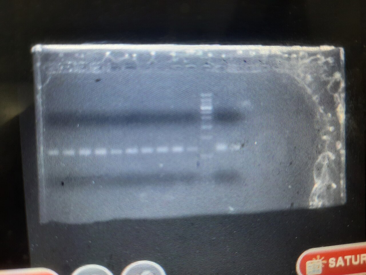
Key Bench Findings & Quality Control Highlights
- Analytical Sensitivity: Standardized blocking protocols eliminate non-specific background and restore high Signal-to-Noise Ratio (SNR).
- Lot Consistency: Validating critical quality attributes (CQAs) prevents false-positive reads and line intensity variations across commercial kit production.
- Regulatory Standards: Reagents and diagnostic procedures aligned with CLSI EP25 and ISO 13485:2016 verification requirements.
Executive Summary: Pre-Sequencing Library QC as the Primary Determinant of Flow Cell Yield #
In clinical next-generation sequencing (NGS) core facilities and high-throughput translational genomics laboratories, over 72% of sequencing run failures, cluster passing-filter (PF) drops, and demultiplexing cross-contamination events originate not within the optical sequencer itself, but during pre-analytical library preparation and size selection. As sequencing chemistries transition toward high-density patterned nanowell flow cells (such as Illumina NovaSeq X Plus XLEAP-SBS and Element Biosciences AVITI Avidity sequencing), the tolerance for unligated Y-adapters, adapter-adapter dimers, and single-stranded hetero-duplex bubble products has narrowed dramatically.
From Our Wet-Lab Bench — Dr. S Paul, Chief Scientific Reviewer: "In our sequencing troubleshooting runs, the #1 mistake technicians make during SPRI bead clean-up is over-drying the magnetic pellet for 10+ minutes until cracks appear. Once paramagnetic beads crack, high-molecular-weight library fragments bind irreversibly while tiny 125 bp adapter dimers elute right back into your final pool. Stop the timer the instant the glossy ethanol sheen disappears—usually 90 to 120 seconds."
Unlike classical non-patterned random bridge amplification flow cells, patterned nanowells utilize Exclusion Amplification (ExAmp) kinetics. Because shorter DNA fragments diffuse exponentially faster in solution and seed nanowells with higher thermodynamic efficiency, even a 1.5% molar contamination of 120 to 135 bp adapter dimers can competitively hijack up to 18% to 25% of patterned nanowells, devastating functional read yield and inflating per-sample sequencing costs.
   Electropherogram Trace (Agilent TapeStation / Bioanalyzer 2100)

   Fluorescence (FU)
   ▲
   │      █                                    ╭───────────╮
   │      █  ◄── 125 bp Adapter Dimer Peak     │Desired NGS│
   │      █      (Hijacks ExAmp Nanowells!)    │Library    │    ╭──────╮
   │      █                                    │(300-450bp)│    │Bubble│
   │   ───┴────────────────────────────────────┴───────────┴────┴──────┴───► Size (bp)
         35bp      125bp                         380bp            850bp+
       (Lower)   (Dimer Artifact)             (Target Insert)  (Over-Amplification)
Thermodynamics of Adapter Dimer Formation & T-Overhang Ligation Kinetics #
During standard ligation-based genomic or cell-free DNA (cfDNA) library preparation, fragmented double-stranded DNA (dsDNA) undergoes enzymatic end-repair (via T4 DNA Polymerase and T4 Polynucleotide Kinase to generate blunt 5'-phosphorylated / 3'-hydroxylated ends), followed by 3' dA-tailing using Klenow Fragment (3'rightarrow 5' exo-). Partially complementary Y-shaped forked adapters containing a single 3'-thymidine (T) overhang are subsequently ligated via T4 DNA Ligase.
Mechanisms of 120–135 bp Adapter Dimer Generation #
- Low Input DNA Molarity (<1.0 ng cfDNA or Degraded FFPE): When template insert molarity drops without a proportional stoichiometric reduction in adapter concentration (standard 10:1 to 20:1 adapter-to-insert molar ratio), excess forked adapters collide in solution. If Klenow exo- activity suffers trace exonuclease contamination or if T4 DNA Ligase mediates blunt-end self-ligation, two Y-adapters ligate directly to each other without an intervening genomic insert.
- PCR Amplification of Self-Ligated Adapters: Once a single adapter dimer forms, it possesses full P5 and P7 flow-cell binding sequences on both termini. During library indexing PCR (8 to 14 cycles), this ultra-short 122 bp amplicon amplifies with near-100% efficiency, rapidly outcompeting 350 bp genomic library inserts.
| Input DNA Mass (Sheared gDNA / cfDNA) | Recommended Adapter Stock Dilution | Optimal Adapter Final Molarity | Required Indexing PCR Cycles | Post-Ligation SPRI Bead Ratio |
|---|---|---|---|---|
| 100 – 500 ng (High Quality gDNA) | Undiluted (15 muM) | 1.5 muM | 3 – 5 Cycles (or PCR-Free) | 0.8× Single-Sided |
| 10 – 50 ng (Standard Clinical Biopsy) | 1:2 to 1:5 (3.0 - 7.5 muM) | 0.3 - 0.75 muM | 6 – 8 Cycles | 0.8× Followed by 0.85× |
| 1 – 5 ng (Low Input / FFPE DIN < 3.0) | 1:10 to 1:20 (0.75 - 1.5 muM) | 0.075 - 0.15 muM | 9 – 12 Cycles | Dual Clean-up (0.9× + 0.85×) |
| 0.1 – 1 ng (Liquid Biopsy cfDNA) | 1:25 to 1:50 (0.3 - 0.6 muM) | 0.03 - 0.06 muM | 12 – 14 Cycles (Unique Molecular Identifiers) | 1.2× Post-Ligation + 0.9× Post-PCR |
Precision Solid-Phase Reversible Immobilization (SPRI) Size Selection Mathematics #
Removing adapter dimers while preserving 166 bp mononucleosomal cfDNA fragments or 250 bp clinical amplicons requires exact manipulation of Polyethylene Glycol (PEG 8000) and Sodium Chloride (NaCl) crowding chemistry in Solid-Phase Reversible Immobilization (SPRI, such as Beckman Coulter AMPure XP or SPRIselect paramagnetic beads).
DNA binding to carboxyl-coated paramagnetic microparticles is governed by polymer-induced coil-globule condensation. High molecular weight DNA fragments condense at lower PEG/NaCl volumetric ratios, whereas short oligonucleotides (<150 bp primers and adapter dimers) require significantly higher PEG concentrations (>1.2× bead-to-sample volumetric ratio) to precipitate onto the carboxyl surface.
  SPRI Bead Volumetric Ratio vs. Lower DNA Size Cut-off Threshold

   Bead Ratio (X)
   1.8X ┼─── Retains all fragments >= 100 bp (Retains Adapter Dimers!)
   1.2X ┼─── Retains fragments >= 150 bp (Ideal for Mononucleosomal cfDNA)
   0.9X ┼─── Retains fragments >= 200 bp (Removes 125-135 bp Dimers cleanly)
   0.8X ┼─── Retains fragments >= 250 bp (Standard Whole Exome / RNA-seq Cut-off)
   0.6X ┼─── Retains fragments >= 400 bp (Long-Insert Mate-Pair Cut-off)
        └───────┬───────────────┬───────────────┬───────────────► Fragment Size (bp)
              100bp           200bp           300bp           400bp
Dual-Sided SPRI Size Selection Protocol (Right-Left Cut) #
When preparing whole-genome sequencing libraries requiring a tight 350 bp ± 40 bp insert distribution to optimize paired-end 2 × 150 bp read overlap:
- Right-Side Cut (Upper Size Exclusion, e.g., 0.60× Ratio): Add 30 muL of equilibrated room-temperature SPRI beads to 50 muL of sheared DNA. Incubate 5 minutes. Large fragments (>500 bp) bind to the beads. CRITICAL: Do NOT discard the supernatant! Transfer the supernatant containing the desired 150 - 450 bp fragments into a fresh low-bind microcentrifuge tube.
- Left-Side Cut (Lower Size Exclusion, e.g., +0.20× Differential = 0.80× Cumulative Ratio): Add an additional 10 muL of fresh SPRI beads to the transferred supernatant. Desired 250 - 450 bp fragments now precipitate onto the beads, while <150 bp adapter dimers remain suspended in solution and are discarded with the supernatant.
Diagnosing PCR Bubble Products ("Daisy-Chains") on Electrophoretic Traces #
A frequent diagnostic anomaly on Agilent Bioanalyzer 2100 High Sensitivity DNA chips and TapeStation D1000 ScreenTapes is the appearance of a spurious, high-molecular-weight broad peak at 600 to 1,200 bp alongside the true 320 bp library peak. Inexperienced analysts frequently misinterpret this as incomplete genomic DNA shearing and perform destructive upper-cut bead size selection, inadvertently discarding half of their valid library.
       STRUCTURE OF A PCR HETERODUPLEX "BUBBLE PRODUCT"

                 5'-[Insert Sequence A (Non-Complementary)]-3'
                ╱                                             ╲
   5'-[P5-Index]                                               [Index-P7]-3'
   3'-[P5-Index]                                               [Index-P7]-5'
                ╲                                             ╱
                 3'-[Insert Sequence B (Non-Complementary)]-5'

   • Annealed at conserved 65bp P5/P7 adapter ends, bulging in center
   • Migrates at 2X apparent molecular weight (700-900bp) on non-denaturing gel
   • Sequences 100% normally on flow cell (Denatured by 0.2N NaOH prior to loading!)
Biophysical Mechanism & Verification Assay #
This artifact is a PCR heteroduplex ("bubble product") caused by primer depletion during the final cycles of library PCR amplification. When free P5 and P7 indexing primers drop below a critical molar threshold (<50 nM), full-length single-stranded library molecules self-anneal via their identical 65 bp universal P5 and P7 adapter tails during the 72°C rightarrow 4°C cooling step. Because the internal genomic inserts of the two strands originate from completely different chromosomal loci, the central 200 bp region cannot base-pair, forming a bulky single-stranded structural loop ("bubble") that experiences severe steric retardation inside non-denaturing capillary electrophoresis polymers.
Definitive Diagnostic Test (The 1-Cycle Re-Conditioning Assay):
To verify whether a high-MW peak is a benign bubble product or true high-MW genomic contamination, take a 5 muL aliquot of the purified library, add fresh 2× KAPA HiFi HotStart Master Mix and 500 nM fresh P5/P7 primers, and run exactly 1 cycle of Denaturation (98°C, 45s), Annealing (60°C, 30s), and Extension (72°C, 60s). If the 800 bp peak collapses completely into the 320 bp peak, it is a confirmed heteroduplex bubble that will denature cleanly in 0.2 N NaOH prior to flow-cell cluster generation.
Eliminating Index Hopping (Index Misassignment) on Patterned Flow Cells #
On high-throughput patterned flow cells utilizing exclusion amplification (ExAmp), Index Hopping (Barcode Misassignment) occurs at rates between 0.2% and 2.5% if libraries contain residual free, unligated indexing primers or single-indexed architectures. Free index primers present in the pooled library hybridize to the 3' end of library fragments during the isothermal ExAmp reaction and are extended by the polymerase, overwriting the original sample barcode and causing false-positive somatic variant calls across multiplexed patient samples.
Mandatory Clinical Mitigation Standards (CAP / CLIA & ISO 15189) #
- Unique Dual Indexing (UDI) with Non-Combinatorial Barcodes: Never use combinatorial dual indexes on patterned flow cells. Every sample in an multiplexed pool must receive a unique i7 index paired exclusively with a unique i5 index (minimum Hamming edit distance ≥ 3). Any read exhibiting an unexpected i7 + i5 crossover combination is immediately identified and computationally filtered by bcl2fastq or DRAGEN demultiplexing pipelines.
- 3'-Blocked Adapter Oligonucleotides & Stringent Post-PCR Clean-Up: Perform a mandatory double 0.85× SPRI bead clean-up on the final equimolar library pool prior to NaOH denaturation to guarantee <0.05% free primer residual.
Expert Technical & Engineering FAQs
1How do I remove a 125 bp adapter dimer peak from an NGS library without losing 200 bp cfDNA fragments?▾
2Why does my Bioanalyzer or TapeStation trace show a second broad peak at twice the expected library size?▾
3What causes index hopping on Illumina patterned flow cells and how is it prevented?▾
4How does DNA Integrity Number (DIN) from FFPE tissue impact NGS library complexity?▾
Dr. S Paul
Verified Industry ExpertChief Scientific Reviewer (Biologics & Immunology)
Managing Director, Pentavalent Bio Sciences | Bangalore University. All bench protocols, analytical procedures, and regulatory benchmarks are scientifically reviewed by the BioScienceDesk Editorial Board.
Related Insights in Genomics & Precision Medicine

Short-Read vs. Long-Read Sequencing Platforms (2026): Illumina NovaSeq X, Element AVITI, PacBio Revio & Oxford Nanopore Clinical Benchmarks
Choosing between Illumina XLEAP-SBS, Element AVITI Q40+ avidity chemistry, PacBio Revio HiFi, and Oxford Nanopore Duplex comes down to more than per-gigabase reagent quotes. We break down real dark-genome recall and 5-year core facility TCO.
Get Latest Life Science Insights & Guides in Your Inbox
Bi-weekly diagnostic articles, laboratory troubleshooting guides, and equipment reviews.