The Day-Eight Panic: Batch Non-Sterility or Laboratory Artifact? #
Look, if you work in an aseptic cleanroom or run batch-release microbiology, you dread day eight.
You walk into the 32-degree incubator room at 7:30 in the morning, shine your inspection light across the stainless racks, and there it is. Canister B has a faint, milky swirl sitting right above the membrane.
Your stomach drops. You have a four-hundred-thousand-dollar lot of sterile injectable vials sitting in quarantine, and QA is already asking for release paperwork.
Hold on before you panic and notify quality assurance. Take a close look at historical fill-finish data. Over two-thirds of positive canisters turn out to be bench accidents. Maybe a tech grazed the manifold port with a sterile sleeve. Maybe an exhaust vent filter got wet during priming. Or maybe someone forgot to rinse out an antimicrobial excipient before adding the growth medium.
Differentiating a real batch failure from a lab mishap comes down to membrane physics, broth chemistry, and how fast you track down the bug.
Filtering vs Direct Inoculation: The Dirty Secret #
USP <71> gives you two choices. If your drug dissolves in water or oil, you filter it. Direct inoculation is an absolute nightmare unless you have no other choice.
Why? Simple physics: you cannot see through it. Put five milliliters of a thick ophthalmic suspension or oily vehicle directly into clear thioglycollate broth, and the whole tube goes cloudy on day zero.
Try telling an FDA auditor on day seven whether that cloudiness is bacteria dividing or just chemical precipitation. You can't. You have to subculture a sample into fresh broth for another four days, which just gives your analyst a second chance to accidentally sneeze a microbe into the tube.
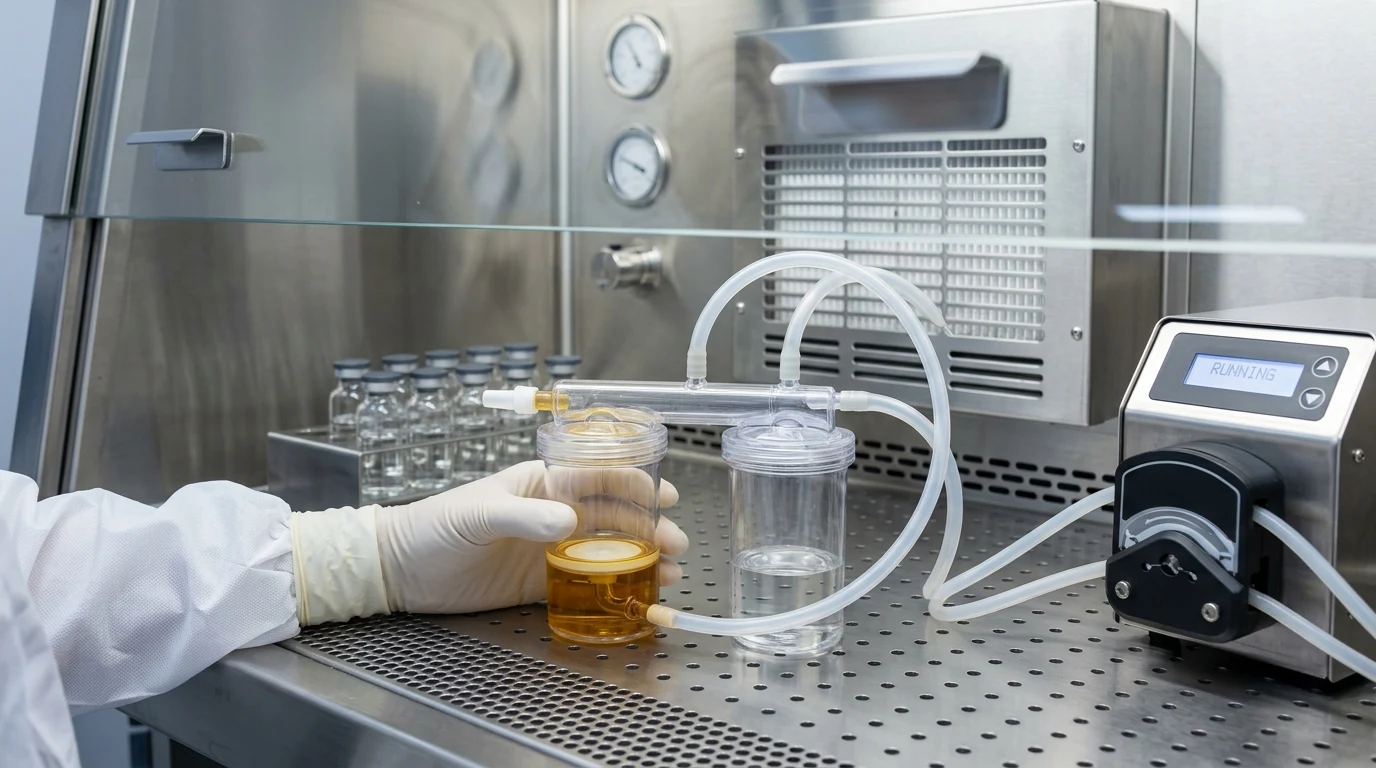
With membrane filtration, you avoid that trap. You pull your pooled vials through twin 0.45-micron hydrophilic PVDF canisters (or mixed cellulose esters for small molecules), catch whatever is swimming in the liquid on the filter face, and then you wash it.
That rinse step is everything. If your formulation has benzyl alcohol, phenol, or an active antibiotic, those chemicals will kill off trace contaminants in the broth and mask a real problem. Flushing the membrane with two or three 100 mL slugs of Fluid A (peptone water) or Fluid D (with 0.1% Polysorbate 80) washes the drug straight into the waste carboy while keeping any live bacteria stuck on the filter.
Method Comparison: Membrane Filtration vs. Direct Inoculation #
| Analytical Parameter | Closed Membrane Filtration (Canisters) | Direct Inoculation (Tubes / Bottles) |
|---|---|---|
| Primary Indication | Aqueous solutions, filterable oils, reconstituted dry powders, biologics | Opaque suspensions, surgical hydrogels, medical fabrics, cell therapies |
| Sample Volume Handled | Full composite lot pools (up to 500 mL – 1,000 mL per manifold) | Restricted sample aliquots (typically ≤ 10% of total broth volume) |
| Microbial Concentration | Concentrates trace bioburden across entire batch onto 0.45 µm disc | Dilutes trace cells into large broth volume, lowering detection probability |
| Inhibitory Drug Washout | Up to 5 × 100 mL rinses with Fluid A, D, or K removes all active excipients | Active drug remains in broth for all 14 days, requiring enzymatic neutralizers |
| Turbidity Interference | Zero broth turbidity; broth fills pre-rinsed, crystal-clear canisters | High turbidity on Day 0; requires secondary 4-day transfer subculturing |
| False-Positive Risk | Lowest (< 0.2% in isolator systems due to closed fluidics) | High (open-vial transfers increase manipulation contact points) |
What About the Two Media Broths? #
USP <71> makes you incubate two formulations for two full weeks.
First is Fluid Thioglycollate Medium, or FTM. We keep this chamber set between 30 and 35 degrees Celsius because that is where common bacterial contaminants thrive—bugs like Staphylococcus aureus, Pseudomonas aeruginosa, and anaerobic Clostridium sporogenes. The broth has sodium thioglycollate and L-cystine mixed in to scavenge dissolved oxygen, keeping the bottom portion anaerobic. At the top, resazurin acts as an oxygen flag. If you see the pink band spread down more than twenty percent of the liquid column, stop. That means oxygen penetrated too deep, and anaerobes will not survive. You have to discard that unit or reheat it to drive off the dissolved air before you load samples.
Second is Soybean-Casein Digest Medium, which most techs call TSB. That one sits at a cooler 20 to 25 degrees Celsius for fourteen days. We keep it cool intentionally so slow-growing yeasts like Candida albicans and fungal spores like Aspergillus brasiliensis do not burn out before forming visible fuzzy colonies.
Method Suitability: The Stasis Test Most Labs Flunk #
Now comes method suitability, the step where half of new drug filings run into trouble.
You cannot just take a new monoclonal antibody or suspension, pump it through two canisters, and claim you verified sterility. You have to prove the drug itself does not suppress microbial growth on the filter. In the old pharmacopeias, everyone called this the bacteriostasis and fungistasis test.
The protocol sounds simple enough: you inoculate fewer than one hundred colony units of six reference bugs directly into your final wash fluid or broth. You need to see clear turbidity within three to five days. If your drug sticks to the filter face and kills the challenge bugs, your test is completely dead in the water.
How to Fix Formulation Kill on the Membrane #
If the drug has parabens or oily preservatives, standard peptone water—Fluid A—will not cut it. We switch immediately to Fluid D, which adds one gram per liter of Polysorbate 80. That tiny bit of detergent breaks the hydrophobic grip on the filter and flushes the preservative out into the drain carboy. If we are testing an ointment or oily vehicle, we step up to Fluid K, which contains beef extract and a heavier peptone blend. And for beta-lactams or cephalosporins, you do not even try washing them out with buffer alone; you add sterile beta-lactamase enzyme right into the rinse line so it chews up the antibiotic ring before the media touches the membrane.
When a Canister Turns Cloudy: The Bench SOP #
When a canister does turn cloudy on day eight, what is the actual investigative protocol on the floor?
Do not agitate the canister. The second you shake it, you destroy the colony morphology and aerate the thioglycollate. Instead, draw one milliliter with a sterile needle and run a rapid Gram stain while streaking an isolate onto blood agar for MALDI-TOF identification.
If the mass spec hits Cutibacterium acnes, Micrococcus luteus, or coagulase-negative Staph, look straight at gowning practice. Those are human skin commensals. You pull the environmental monitoring plates from that day's run and check the operator's fingertip touch plates. If the identical strain pops up on the analyst's right glove, you have hard evidence of a lab breach.
On the other hand, if you pull Ralstonia pickettii or Burkholderia cepacia, stop looking at the analyst. Those are waterborne Gram-negative rods. That points straight back to your cleanroom water loop or an autoclave condensation issue during vial washing.
Under FDA guidance and EU Annex 1, you cannot retest a positive lot just because you feel lucky. Unless you have definitive, documented lab error—like a breached isolator glove or a contaminated negative control—that batch is rejected.