Beyond Glucose: The Dermal Interstitial Fluid Frontier #
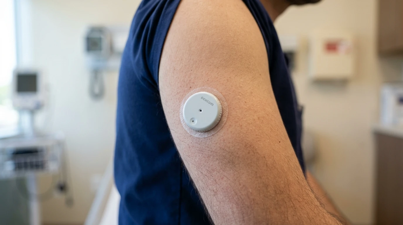
If you listen to consumer wearable keynotes, you would think the holy grail of health tracking begins and ends with continuous glucose monitoring.
Dexcom and Abbott built forty-billion-dollar empires on glucose because glucose was the easiest biochemical target in human physiology. It circulates at comfortable millimolar concentrations (4 to 10 mM), and nature gave us glucose oxidase—an unusually rugged, high-turnover enzyme that survives for two weeks on a subcutaneous wire without losing activity.
Step outside glucose into clinical critical care, oncology, or endocrinology, and glucose oxidase tricks stop working.
Doctors in intensive care units do not manage septic shock by guessing glucose curves; they titrate vasopressors against blood lactate. Oncologists and transplant surgeons dosing vancomycin or tacrolimus walk a razor-thin line between therapeutic efficacy and irreversible nephrotoxicity, relying on single-point trough blood draws taken hours too late.
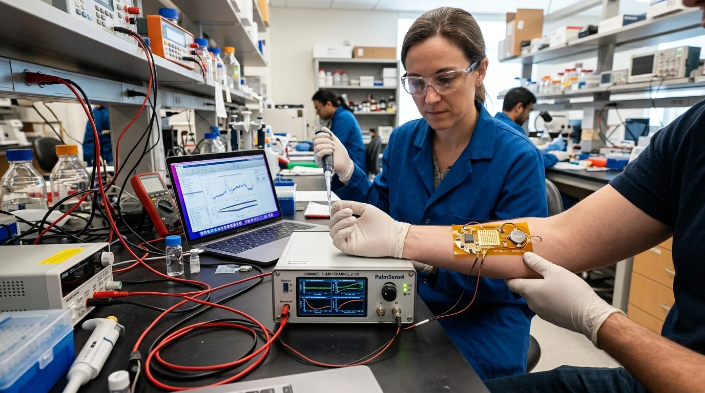
The true diagnostic frontier is continuous multi-analyte monitoring in dermal interstitial fluid (ISF) using microneedle arrays.
Why Dermal Interstitial Fluid Beats Blood Draws #
Dermal interstitial fluid is the biochemical bath surrounding living cells in the epidermis and dermis. Because capillary walls are semi-permeable, small-molecule metabolites and unbound therapeutic drugs equilibrate rapidly between intravascular blood and dermal ISF.
For decades, accessing ISF required painful microdialysis catheters or vacuum blister extraction that took hours to collect five microliters.
Microneedle biosensors bypass that barrier mechanically. An array of micro-machined projections—typically 500 to 800 microns in length—penetrates through the dead, keratinized stratum corneum into the viable epidermis. Because the needles terminate above the deep dermal capillary plexus and pain-sensing nociceptive nerve endings, patch insertion is completely painless and draws zero capillary blood.
Microneedle Architecture Tradeoffs in Interstitial Fluid Sensing #
| Engineering Architecture | Hollow Microneedles (Aspiration) | Swellable Hydrogel Arrays (Extraction) | Solid Coated Arrays (In Situ Electrochemistry) |
|---|---|---|---|
| Sensing Mechanism | External electrochemical cell fed by fluid aspiration | Post-wear chemical desorption from swollen polymer | Direct in situ amperometry / voltammetry at needle tip |
| Response Latency | 5 to 15 minutes (fluidic transport delay through micro-lumen) | Off-line analysis only (hours to days post-patch removal) | Sub-second (< 2 seconds electrochemical Faradaic response) |
| Clogging Liabilities | High (keratin plugs, dermal tissue compression, air bubbles) | N/A (hydrogel acts as fluid sink via capillary osmotic swelling) | Zero fluidic clogging (no lumens, no microfluidic channels) |
| Multiplexing Capability | Difficult (requires multi-channel microfluidic manifolds) | Moderate (limited by spatial resolution during extraction) | High (individual micro-posts functionalized with distinct bioreceptors) |
| Clinical Target Application | High-volume drug delivery / insulin infusion systems | Pharmacokinetic mass spectrometry research studies | Continuous real-time critical care telemetry & wearable TDM |
Solid Coated Arrays vs Hollow Microfluidics #
Bioengineers have explored three distinct microneedle architectures over the past decade: hollow, swellable hydrogel, and solid coated arrays.
Hollow microneedles act like miniature hypodermic needles that aspirate ISF into an external sensor chamber. In practice, hollow needles clog constantly. Keratin debris and dermal tissue compression plug the 50-micron lumens during insertion, while sluggish capillary flow introduces ten-minute microfluidic transport lags.
Swellable hydrogel arrays absorb interstitial fluid like a sponge, but extracting the captured analyte requires post-wear centrifugation, making real-time continuous telemetry impossible.
The winning clinical architecture is the solid electrochemical microneedle array. Here, you do not extract fluid at all. You turn the microneedle surface itself into an electrode:
The array packs working, reference, and counter electrodes on adjacent micro-posts. Sensing occurs directly in situ within the dermal fluid bath, delivering sub-second response kinetics.
The Transduction Shift: From Enzymes to Structure-Switching Aptamers #
For continuous lactate, enzymatic sensing still holds up. We immobilize lactate oxidase onto platinum-coated microneedles, protecting the surface with an inner permselective layer of poly-m-phenylenediamine to block ascorbic acid and acetaminophen interference.
When you shift from lactate to cortisol or therapeutic drug monitoring, however, nature runs out of oxidases. There is no simple oxidase enzyme for vancomycin, methotrexate, or doxorubicin.
To track non-enzymatic targets continuously, the field relies on Electrochemical Aptamer-Based (E-AB) sensors.
How E-AB Transduction Works on the Microneedle Tip #
You self-assemble a dense monolayer of synthetic single-stranded DNA aptamers onto a gold microneedle surface. The distal end of each aptamer strand is tagged with a methylene blue redox reporter.
- Unbound Conformation: In the absence of target drug, the flexible DNA strand keeps the methylene blue tag in rapid electronic contact with the gold electrode, generating high Faradaic current.
- Target Binding Fold: When the target drug (like vancomycin) binds specifically to the aptamer pocket, the DNA strand undergoes a reversible conformational reorganization that shifts the methylene blue tag further away from the gold surface, slowing electron transfer kinetics.
- Reversible Equilibrium: By running continuous square-wave voltammetry, the biosensor measures the change in Faradaic peak current. Because binding is non-covalent and completely reversible, when local drug concentration drops, the drug dissociates, the aptamer resets, and current recovers within seconds.
The 48-Hour Dermal Biofouling Barrier #
The hardest problem in wearable microneedle biosensing is not electronic sensitivity; it is biofouling.
Within minutes of penetrating the skin, non-specific protein adsorption coats the electrode surface with albumin and fibrinogen. Over the next twelve to twenty-four hours, the body mounts an acute local foreign-body response, forming a fibrous protein capsule that restricts diffusion and causes sensor signal drift.
Overcoming sensor drift requires outer antifouling membrane engineering. Commercial-grade patches coat microneedle surfaces with zwitterionic poly-carboxybetaine hydrogels or semi-permeable polyurethane membranes. These hydrophilic polymer brushes create a permanent hydration shell of water molecules that physically repels non-specific protein adsorption, extending stable in vivo calibration lifetimes past seventy-two hours.