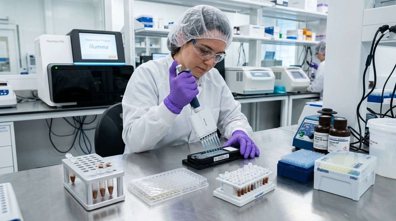
The Archival Pathology Dilemma: Low Input and Damaged Templates #
Every molecular pathologist and NGS lab director knows the sinking feeling when a clinical oncology biopsy arrives from pathology archives.
It is a paraffin block from 2019. The tumor tissue inside is an eight-millimeter needle core from a non-small cell lung cancer patient. The medical oncologist wants comprehensive genomic profiling across 500 genes to hunt for rare EGFR exon 20 insertions and KRAS G12C mutations. And your histology tech hands you three microtome curls in a 1.5 mL tube.
If you treat that formalin-fixed tissue like fresh frozen cell culture, your sequencing run is doomed before you even touch the library prep bench.
Formalin fixation preserves tissue morphology for microscopic staining, but it is an absolute nightmare for nucleic acids. Neutral buffered formalin reacts with free amino groups on lysine residues and nucleotide bases, forming stable methylene bridges (-CH2-) between histone proteins and DNA strands. Over months and years in storage, ambient moisture drives hydrolytic deamination of cytosine into uracil, while tissue hypoxia causes extensive enzymatic shearing.
Extracting sequencing-grade DNA and RNA from archival FFPE requires three strict biochemical controls: complete deparaffinization without organic carryover, controlled thermal uncoupling of cross-links, and enzymatic removal of deaminated bases before PCR amplification.
Deparaffinization: Why Xylene Is Being Replaced on the Bench #
For decades, the standard laboratory protocol relied on toxic xylene washes followed by sequential ethanol rehydration (100%, 95%, 70%).
While xylene dissolves paraffin effectively, it introduces two serious failure modes:
- Enzyme Inhibition: Trace residual xylene or ethanol carried through the wash steps poisons proteinase K digestion and downstream polymerases.
- Physical Pellet Loss: Centrifugal pelleting of brittle, dehydrated tissue curls often causes sample loss on tube walls, particularly with low-cellularity core biopsies.
Modern clinical pipelines rely on enzymatic or non-toxic mineral oil partitioning. Commercial deparaffinization solutions sit on top of the aqueous lysis buffer during incubation. As the heat block ramps to 56 degrees Celsius, the paraffin melts and partitions into the upper organic phase, allowing proteinase K to access tissue fragments directly in the lower aqueous buffer without centrifugation.
Deparaffinization & Extraction Chemistry Benchmarks #
| Extraction Step | Classical Xylene / Ethanol Method | Modern Mineral Oil / Aqueous Phase Partitioning |
|---|---|---|
| Paraffin Removal Mechanism | Chemical solvent dissolution via aromatic hydrocarbon washes | Thermal phase partitioning (paraffin melts into top organic layer) |
| Hands-On Bench Time | 45 to 60 minutes (multiple centrifugation and aspiration cycles) | 5 minutes (single-tube addition, zero pre-lysis centrifugation) |
| Low-Input Core Biopsy Recovery | Poor (< 50% recovery due to pellet fragmentation and wall sticking) | High (> 90% recovery; tissue remains submerged in aqueous lysis buffer) |
| Residual Solvent Hazard | High risk of ethanol/xylene carryover poisoning PCR polymerases | Zero organic solvent carryover into magnetic bead bind step |
| Fume Hood Requirement | Mandatory chemical fume hood for toxic aromatic vapors | Standard molecular biology benchtop or biosafety cabinet |
The Cross-Link Reversal Dilemma: DNA Heat vs RNA Fragility #
Once tissue is rehydrated, you must break the methylene cross-links holding proteins to the DNA backbone. This requires heat, but heat is a double-edged sword.
For genomic DNA, high-temperature incubation at 90 degrees Celsius for 60 to 90 minutes is essential to hydrolyze methylene bridges. Without this thermal step, intact histones block polymerase extension during NGS target enrichment.
For total RNA, however, 90 degrees Celsius is catastrophic. Heat in the presence of trace divalent cations triggers rapid phosphodiester backbone hydrolysis, reducing your already damaged RNA into twenty-nucleotide fragments. Clinical FFPE RNA extraction protocols cap thermal uncoupling at 56 to 65 degrees Celsius, relying on prolonged proteinase K digestion (often overnight at 56°C) rather than aggressive boiling.
The C>T Deamination Artifact: Eliminating False Somatic Mutations #
The most dangerous artifact in FFPE oncology sequencing is hydrolytic cytosine deamination.
Over time, water molecules in formalin-fixed blocks convert cytosine into uracil:
During NGS library construction, high-fidelity proofreading DNA polymerases read uracil as thymine. On the complementary strand, adenine is inserted instead of guanine. The result is a flood of artificial C>T and G>A transition artifacts.
In clinical liquid biopsy or tissue panels hunting for low-frequency subclonal mutations (1% to 3% variant allele frequency), these deamination artifacts look identical to true oncogenic driver mutations—particularly in CpG hotspots of KRAS and TP53.
How to Eliminate Deamination Noise #
Before fragmentation or adapter ligation, you treat the extracted DNA with Uracil-DNA Glycosylase (UDG). UDG specifically cleaves the uracil base from the deoxyribose phosphate backbone, creating an abasic site. When the template enters PCR, heat breaks the phosphodiester chain at the abasic site, preventing the artifactual strand from amplifying and preserving only authentic genomic DNA for sequencing.
Quality Control Metrics: Why NanoDrop Must Be Banned for FFPE #
Never rely on a NanoDrop UV spectrophotometer (A260/A280) to quantify FFPE DNA.
Formalin-fixed extracts contain substantial amounts of free nucleotides, hydrolyzed RNA fragments, and cellular protein debris. NanoDrop absorbs light at 260 nm regardless of whether the molecule is intact double-stranded DNA or degraded single nucleotides, routinely overestimating concentration by two- to five-fold.
Always quantify FFPE yields using fluorometric dye binding (Qubit dsDNA High Sensitivity assay), which binds specifically to double-stranded DNA.
Integrity Assessment: DIN and DV200 #
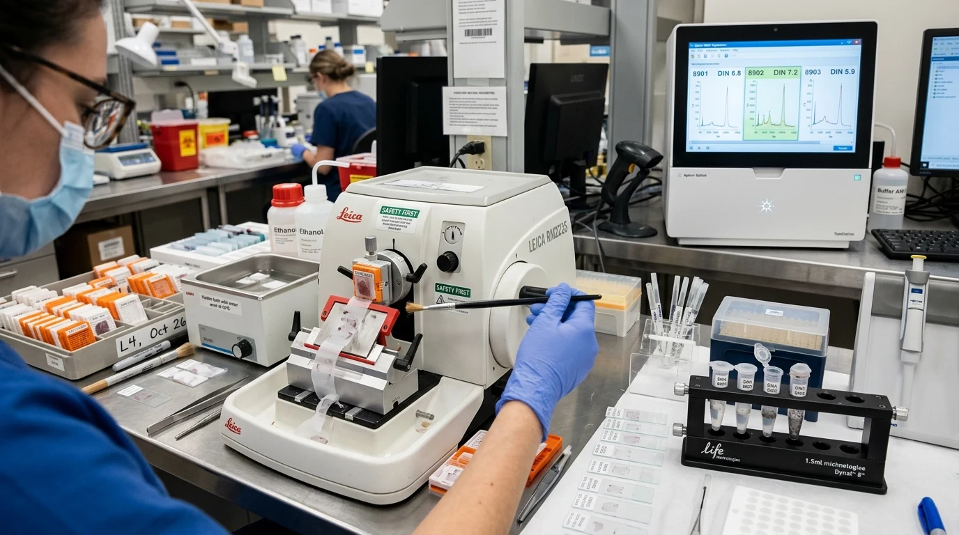
For integrity assessment, run capillary electrophoresis on an Agilent 4200 TapeStation:
- Genomic DNA: Track the DNA Integrity Number (DIN). A DIN score above 6.0 indicates robust genomic DNA suitable for large hybridization-capture panels. For DNA with DIN between 3.0 and 5.0, increase library input mass by 50% to compensate for shorter template length.
- Total RNA: Abandon the classical 28S/18S ribosomal RNA Integrity Number (RIN). Instead, measure the DV200 metric—the percentage of RNA fragments longer than 200 nucleotides. If DV200 exceeds 50%, the sample will reliably generate comprehensive RNA-Seq libraries for oncology fusion detection.